The sacroiliac joint (SIJ) is a vital structure in the human body that plays a central role in stabilizing the pelvis, supporting the weight of the upper body, and facilitating efficient movement during walking and other physical activities.
While often overlooked compared to other major joints, such as the hip or knee, the SIJ is critical in connecting the spine to the pelvis and enabling smooth, coordinated movement.
SIJ injuries can be disabling and require careful diagnosis and treatment, particularly in cases involving fractures or dislocations.
This article aims to provide an in-depth look at the anatomy, function, types of fractures, and associated injuries of the sacroiliac joint, with a focus on recent research and clinical practices.
What is the Sacroiliac Joint (SIJ)?
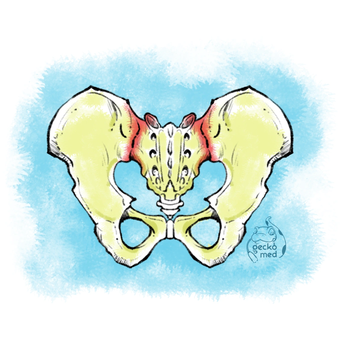
The sacroiliac joint is a synovial joint located at the interface between the sacrum, which is the triangular-shaped bone at the base of the spine, and the ilium, one of the major bones of the pelvis.
There are two sacroiliac joints, one on each side of the sacrum.
These joints play a key role in transferring the weight of the upper body to the lower limbs and maintaining pelvic stability during movement.
The SIJ is formed by the articulation of the sacrum and ilium, where the articular surfaces are covered by cartilage, allowing for limited motion.
Unlike most joints in the body that are designed to allow a wide range of movement, the SIJ has minimal movement.
It primarily serves as a conduit for force transmission rather than a site for extensive mobility.
Its functions, however, are essential for maintaining posture and balance during standing, walking, and running.
Understanding the anatomy of the SIJ is essential for comprehending its function and potential pathologies.
Anatomy of the Sacroiliac Joint
To understand how the SIJ functions and the implications of its injury, it’s important to examine the bones involved, the ligaments that stabilize the joint, and the overall role of the SIJ in human biomechanics.
Bones Involved
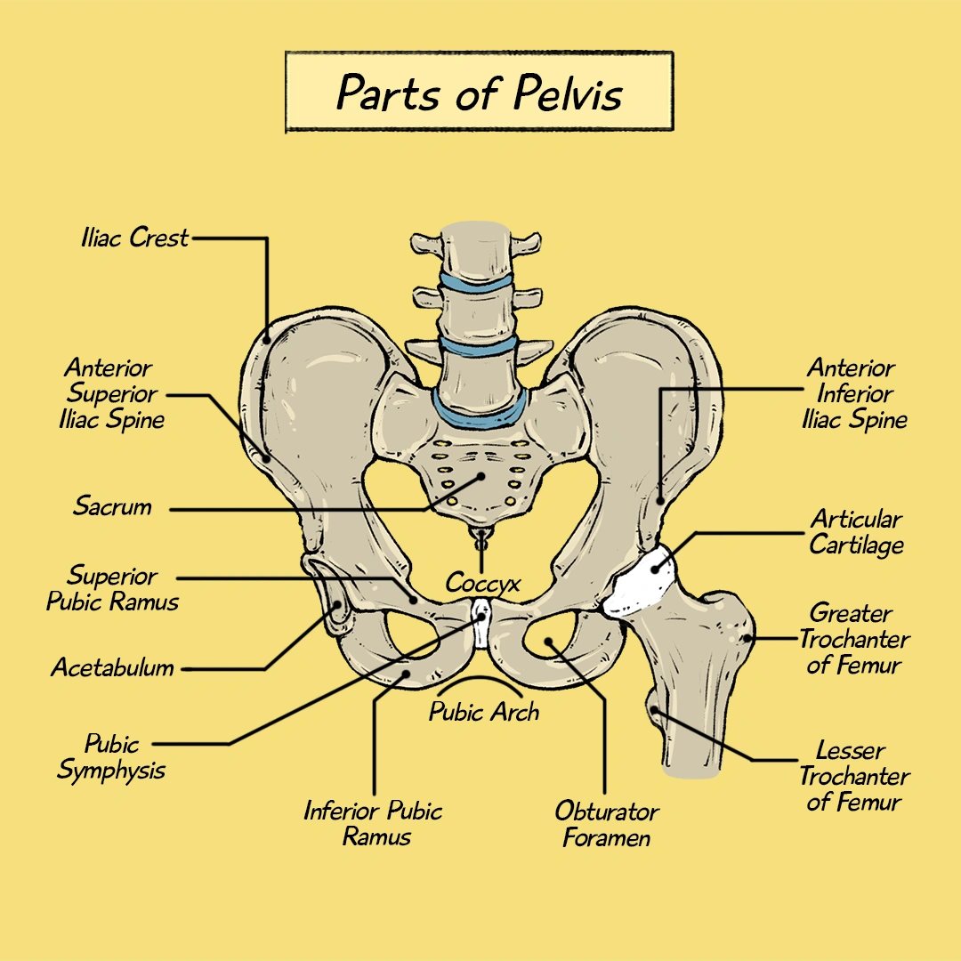
Sacrum: The sacrum is located at the bottom of the vertebral column and consists of five fused vertebrae.
It connects the lumbar spine (L5) to the coccyx and forms the posterior portion of the pelvic ring.
The sacrum has a concave shape that facilitates its connection to the iliac bones.
The sacrum’s surface is rough, which helps it to engage with the iliac bones more securely.
Ilium: The ilium is the largest part of the pelvic bone and forms the upper portion of the hip bone.
Each ilium connects to the sacrum on the left and right sides, creating the sacroiliac joints.
The ilium is broad and fan-shaped, and it plays a crucial role in weight-bearing functions.
Together, these bones form the SIJ on both sides of the pelvis, which is essential for transmitting the weight of the upper body to the lower limbs.
SIJ Functionality
The sacroiliac joint has multiple roles that are crucial for human locomotion and posture:
Weight Bearing: The primary function of the SIJ is to transfer the weight of the upper body to the lower limbs.
When standing, the sacrum transfers the weight from the spine to the ilium and, by extension, to the femur and legs.
This is especially important during activities such as standing and walking.
Pelvic Stability: The SIJ helps stabilize the pelvic girdle during movement, particularly when walking or running.
The joint ensures that the pelvis remains balanced and aligned, which is necessary for efficient and coordinated movement.
Shock Absorption: As the body moves, especially when walking or running, the SIJ helps absorb the shock generated from the ground, reducing the impact on the spine and lower limbs.
Facilitating Motion: While the SIJ does not allow extensive movement, it provides enough motion to accommodate walking, running, and other physical activities.
This limited mobility is important for maintaining flexibility in the pelvis while still providing the stability needed to support movement.
While relatively stable, the SIJ can be susceptible to fractures, often due to high-impact trauma.
Types of Sacroiliac Joint Fractures
Sacroiliac joint fractures are relatively rare compared to fractures of other joints like the hip or knee.
However, when they do occur, they can result in significant discomfort and disability. SIJ fractures are commonly seen in trauma cases, such as falls, motor vehicle accidents, and high-impact sports injuries.
Fractures of the SIJ can be classified into different types, depending on the severity, stability, and whether both joints are involved.
Fracture-Dislocations
A fracture-dislocation of the sacroiliac joint involves both a fracture of the bone and a dislocation of the joint.
This is considered one of the most severe types of SIJ injury because it disrupts both the structural integrity and functional stability of the joint.
Fracture-dislocations are most commonly seen in high-energy trauma, such as motor vehicle accidents or falls from significant heights.
The dislocation occurs when the sacrum and ilium are not only fractured but also displaced from their normal anatomical alignment.
This can lead to a highly unstable pelvis and often results in significant bleeding, nerve damage, and damage to internal organs.
Stable vs. Unstable Fractures
SIJ fractures can also be categorized as stable or unstable, depending on how much displacement has occurred.
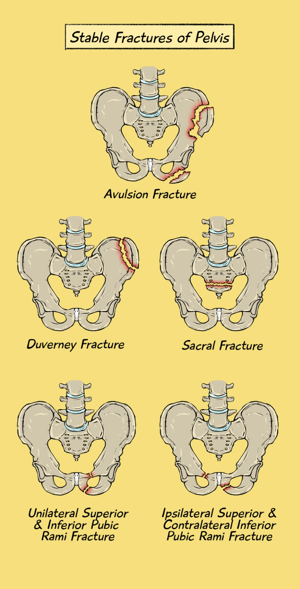
Stable Fractures
Stable fractures of the SIJ involve a break in the bone without significant displacement.
In these cases, the bones remain aligned, and the pelvic ring’s integrity is maintained.
Stable fractures typically result from low-energy trauma and often respond well to conservative management, including rest, pain management, and physiotherapy.
Surgical intervention is generally not required.
Unstable Fractures
Unstable fractures occur when there is significant displacement of the bones, resulting in instability of the pelvis.
These fractures usually require surgical intervention to restore the alignment of the bones and to stabilize the pelvis. In some cases, the instability can lead to nerve damage, pelvic organ injury, or severe bleeding.
Special Types of Unstable Pelvic Fractures
1) Malgaigne Fracture
Mechanism of Injury (MOI): Vertical shear force
Fracture Site:
- Ipsilateral sacroiliac joint disruption
- Ipsilateral superior and inferior pubic ramus fracture
Features: The pelvis becomes vertically unstable.
2) Bucket Handle Fracture
Mechanism of Injury (MOI): Lateral compression from both sides
Fracture Location:
- Ipsilateral sacroiliac joint disruption
- Contralateral superior and inferior pubic ramus fracture
X-Ray Findings: Characteristic “bucket handle” appearance
Features: Horizontal instability of the pelvis.
3) Open Book Fracture
Mechanism of Injury (MOI): Antero-posterior compression
Fracture Location:
- Pubic diastasis (opening up of the pubic symphysis)
- With or without sacroiliac joint disruption
X-Ray Findings: Widening of the pubic symphysis resembling an open book
Features: Horizontal instability of the pelvis.
Unstable fractures are more common in high-energy trauma cases and carry a greater risk of complications.
Bilateral vs. Unilateral Fractures
Another way to classify SIJ fractures is by whether one or both sacroiliac joints are affected.
Bilateral Fractures: Bilateral SIJ fractures involve both sacroiliac joints.
These are considered to be more severe injuries because they result in a significant loss of pelvic stability.
Bilateral fractures are often associated with other pelvic fractures and require immediate surgical intervention to restore alignment and stabilize the pelvic ring.
Unilateral Fractures: Unilateral SIJ fractures affect only one sacroiliac joint.
These fractures are typically less severe than bilateral fractures and can often be managed with conservative treatments.
However, if the fracture is unstable or causes significant pain, surgery may be necessary.
Unilateral fractures are more common and can result from less severe trauma, such as a fall or minor car accident.
Associated Injuries
Sacroiliac joint fractures are often accompanied by other injuries, which can complicate treatment and recovery.
Some of the most common associated injuries include:
Pelvic Ring Fractures: A pelvic ring fracture occurs when there is a break in the bony structures that form the pelvic ring, which includes the sacrum and iliac bones.
These fractures often occur in conjunction with SIJ fractures and can cause significant instability in the pelvic region.
Pelvic ring fractures are associated with a high risk of hemorrhage and may require surgery to stabilize the pelvis.
Soft Tissue and Ligament Damage: The ligaments surrounding the SIJ, such as the anterior and posterior sacroiliac ligaments, can be damaged in traumatic events.
Ligament damage can further destabilize the joint and lead to chronic pain, stiffness, and difficulty walking. Injury to other soft tissues, such as muscles and tendons, can also occur, leading to longer recovery times.
Neurological Damage: In some cases, SIJ fractures, particularly those that involve significant displacement, can cause damage to the nerves that pass through the pelvis.
The sciatic nerve, in particular, may be at risk, leading to pain, numbness, or weakness in the lower extremities.
Internal Organ Injury: High-impact trauma can cause injury to the bladder, intestines, or reproductive organs, which are in close proximity to the sacroiliac joint.
Pelvic fractures, particularly bilateral SIJ fractures, can result in bleeding or perforation of internal organs, which may require emergency intervention.
Understanding the different types of SIJ fractures is crucial for accurate diagnosis and appropriate treatment.
Conclusion
SIJ fractures are complex injuries that require a comprehensive understanding of their anatomy, classification, diagnosis, and management.
Medical students and clinicians involved in the care of these patients should be familiar with the various treatment options, potential complications, and rehabilitation strategies.
By staying abreast of the latest research and advances in the field, healthcare professionals can provide optimal care to patients with SIJ fractures and help them achieve the best possible outcomes.
